Noosa Sports & Spinal Physiotherapy
Leaders in Physiotherapy and Sports Medicine
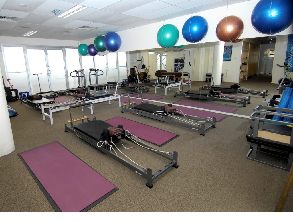
Physiotherapy and allied health services for all ages and stages of life
At Noosa Sports and Spinal Physiotherapy, we provide physiotherapy services to help you get better, feel better and stay better. Our dedicated and experienced team help keep the residents of Noosa, Noosaville, Weyba Downs, Doonan, Noosa Heads, Tewantin healthy and pain free.
-
 (07) 5449 0024
-
 (07) 5449 7774
-
 Suite 202, 90 Goodchap Street
Noosaville, Queensland 4566
-

-

We lead the way when it comes to range, quality and expertise.


Noosa Sports & Spinal Physiotherapy
-
 (07) 5449 0024
-
(07) 5449 7774
-
Suite 202, 90 Goodchap Street
Noosaville, Queensland
4566
-
Open Hours
monday
8:00 am - 6:30 pm
tuesday
7:30 am - 5:30 pm
wednesday
7:30 am - 6:30 pm
thursday
7:30 am - 5:00 pm
friday
7:30 am - 3:30 pm
saturday
Closed
sunday
Closed